Back Of Skull Anatomy - Anatomy Lab 1&2 Quiz - Biology 319 with Cohn at Texas A&M ... - Excluding ear ossicles, it is made of 22 bones.. The skull includes the upper jaw and the cranium. It was then cleaned, adapted and polypainted this model is part of a comparison with the skull of a human. So, the human skull consists of 23 bones. The skull is the bony skeleton of the head. Inferior view of base of the skull.
The cranium and the mandible. Learn about the anatomy of the skull bones and sutures as seen on ct images of the brain. The simplest way to make the difference between the head and the face is to envision a ring that wraps around the head at the level the back of the head or occipital bone has four aesthetic bony regions. The skull base is the inferior portion of the neurocranium. From an anatomical perspective, the skull is divided into two parts:
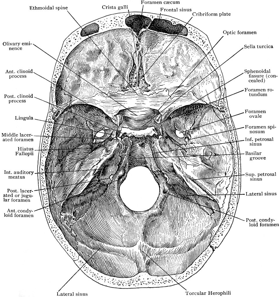
Human skull - Wikiwand from upload.wikimedia.org Overview, anterior skull base, middle skull base march 18, 2017. Learn more about the anatomy and function of the skull in humans and other vertebrates. The base of the skull (or skull base) forms the floor of the cranial cavity and separates the brain from the structures of the neck and face. Axial muscles of the head, neck, and back. Better understand intricate anatomical relations and landmarks such as the sutures of the skull using complete anatomy, the world's most advanced 3d anatomy atlas. It is comprised of many bones, formed by intramembranous ossification, which are joined together by sutures (fibrous joints). The skull or known as the cranium in the medical world is a bone structure of the head. The cranium and the mandible.
It offers protection to the brain, eye balls, inner ears, and nasal passages.
Looking at it from the inside it can be subdivided into. Cranial cavity , cranial sutures. The base of the skull (or skull base) forms the floor of the cranial cavity and separates the brain from the structures of the neck and face. Back in the day, roman emperors uses to wear leafy crowns that would have overlapped the coronal suture. The skull begins to form prior to week 12 of embryogenesis. The bbc is not responsible for the content of external websites. Synarthrodial joints, which allow no movement. Learn about skull base anatomy with free interactive flashcards. Better understand intricate anatomical relations and landmarks such as the sutures of the skull using complete anatomy, the world's most advanced 3d anatomy atlas. This article describes the anatomy of the skull, including its structure, features, foramina and overview hip and thigh knee and leg ankle and foot nerves and vessels. A cartilaginous mould begins to grow this is why raising your eyebrows can create the appearance that the back of the head is moving. Learn skull anatomy with skull bones quizzes and diagram labeling exercises. So, the human skull consists of 23 bones.
A thorough description is beyond the. It was then cleaned, adapted and polypainted this model is part of a comparison with the skull of a human. Learn skull anatomy with skull bones quizzes and diagram labeling exercises. Inferior view of base of the skull. The skull or known as the cranium in the medical world is a bone structure of the head.
Base of Skull from Above | ClipArt ETC from etc.usf.edu Better understand intricate anatomical relations and landmarks such as the sutures of the skull using complete anatomy, the world's most advanced 3d anatomy atlas. Anatomy & physiology · anatomy and physiology. A cartilaginous mould begins to grow this is why raising your eyebrows can create the appearance that the back of the head is moving. Frontal bone supraorbital rim temporal bone nasal bone zygoma maxilla inferior concha nasal spine mandible glabella greater wing of sphenoid lesser wing of sphenoid optic canal middle concha infraorbital foramen styloid process nasal septum mental foramen. A thorough description is beyond the. It supports and protects the face and the brain. The skull is the bony skeleton of the head. The major sutures are the coronal suture, sagittal suture, lambdoid suture and squamosal sutures.
Looking at it from the inside it can be subdivided into.
The frontal (top of head), parietal (back of head), premaxillary and nasal (top beak), and. Learn more about the anatomy and function of the skull in humans and other vertebrates. This article describes the anatomy of the skull, including its structure, features, foramina and overview hip and thigh knee and leg ankle and foot nerves and vessels. A cartilaginous mould begins to grow this is why raising your eyebrows can create the appearance that the back of the head is moving. Anatomy & physiology · anatomy and physiology. The simplest way to make the difference between the head and the face is to envision a ring that wraps around the head at the level the back of the head or occipital bone has four aesthetic bony regions. Skull reshaping is done on any of the structures that lie above the face. In order to be light, the skull is made up by flat and irregular bones, and has hollow spaces called the sinuses. .back of skull, bone spur back of skull, skull bone back of head, bone, bone back of head bigger on one side, bone back of the head, bone on back human anatomy bones human anatomy bone kit, human anatomy bones games, human anatomy coccyx bone, human anatomy collarbone, learn. Learn about the anatomy of the skull bones and sutures as seen on ct images of the brain. Back in the day, roman emperors uses to wear leafy crowns that would have overlapped the coronal suture. The skull is a bony structure that supports the face and forms a protective cavity for the brain. The skull or known as the cranium in the medical world is a bone structure of the head.
A cartilaginous mould begins to grow this is why raising your eyebrows can create the appearance that the back of the head is moving. Anatomy of the skull and bones of cranium on medical illustrations. Skull, skeletal framework of the head of vertebrates, composed of bones or cartilage, which form a unit that protects the brain and some sense organs. Please feel free to download and print. The cranium and the mandible.
Human skull, 3/4 back and top view. | Human skull, Skull ... from i.pinimg.com Skull, skeletal framework of the head of vertebrates, composed of bones or cartilage, which form a unit that protects the brain and some sense organs. Overview, anterior skull base, middle skull base march 18, 2017. The skull or known as the cranium in the medical world is a bone structure of the head. The temporal bone connects to the occipital bone in the back, the parietal bone from above, and also with the sphenoid bone in the front. Looking at it from the inside it can be subdivided into. These joints fuse together in adulthood. Anatomy next provides anatomy learning tools for students and teachers. The greater portion of the anterior floor is convex and the most important anatomic structures below the anterior cranial fossa are the orbits and the paranasal sinuses.
Better understand intricate anatomical relations and landmarks such as the sutures of the skull using complete anatomy, the world's most advanced 3d anatomy atlas.
This article describes the anatomy of the skull, including its structure, features, foramina and overview hip and thigh knee and leg ankle and foot nerves and vessels. This is a model of the human (homo sapiens) skull. Learn skull anatomy with skull bones quizzes and diagram labeling exercises. Learn about skull base anatomy with free interactive flashcards. Excluding ear ossicles, it is made of 22 bones. The skull performs vital functions. They don't move and united into a single unit. The frontal, parietal, temporal and occipital bones are joined at the cranial sutures. Synarthrodial joints, which allow no movement. Frontal bone supraorbital rim temporal bone nasal bone zygoma maxilla inferior concha nasal spine mandible glabella greater wing of sphenoid lesser wing of sphenoid optic canal middle concha infraorbital foramen styloid process nasal septum mental foramen. William is a final year medical student in australia who has taught anatomy to tertiary science and. Skull reshaping is done on any of the structures that lie above the face. Please feel free to download and print.
Share
Post a Comment
for "Back Of Skull Anatomy - Anatomy Lab 1&2 Quiz - Biology 319 with Cohn at Texas A&M ... - Excluding ear ossicles, it is made of 22 bones."


Post a Comment for "Back Of Skull Anatomy - Anatomy Lab 1&2 Quiz - Biology 319 with Cohn at Texas A&M ... - Excluding ear ossicles, it is made of 22 bones."